Spina Bifida Cognitive Risk Calculator
How This Tool Works
This calculator estimates potential cognitive development outcomes based on spina bifida type and key factors. Results provide approximate ranges and should be used as a guide for discussion with healthcare providers.
Select Spina Bifida Type
Additional Factors
Estimated Outcomes
When a pregnancy is screened, Spina bifida is a neural tube defect that occurs when the spine and spinal cord don’t close properly during the first month of gestation. Parents often wonder how this condition, identified before birth, might shape a child’s thinking, learning, and behavior later on.
Key Takeaways
- Spina bifida can affect the brain directly (hydrocephalus, Chiari II malformation) and indirectly through spinal cord injury.
- Early detection via fetal ultrasound and prenatal MRI helps doctors plan interventions.
- Myelomeningocele carries the highest cognitive risk; milder forms like occulta usually have normal intelligence.
- Prenatal surgery can reduce the severity of brain anomalies and improve long‑term learning outcomes, but it’s not a guarantee.
- Multidisciplinary postnatal care, early educational support, and regular neuro‑assessment are critical for maximizing potential.
Understanding Spina Bifida
Spina bifida is part of a broader group called neural tube defects. The defect happens when the embryonic neural tube fails to seal, leaving an opening in the vertebrae. Three main types exist:
- Spina bifida occulta - a hidden defect, often detected incidentally on X‑ray; usually no neurological problems.
- Meningocele - the protective membranes protrude through the spine but the spinal cord stays inside; motor deficits can be mild.
- Myelomeningocele - the spinal cord and nerves protrude; this is the most severe form and the one most linked to cognitive challenges.
While each form mainly concerns the spine, the brain can be affected because the same embryological process shapes both the spinal canal and the posterior fossa.
How Spina Bifida Is Detected Before Birth
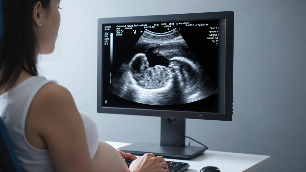
Screening starts with a standard second‑trimester fetal ultrasound. A clear “lemon sign” in the skull or a “banana sign” in the cerebellum points to Chiari II malformation, a brain abnormality commonly seen with myelomeningocele.
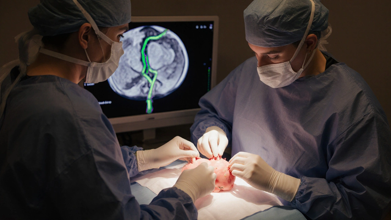
If ultrasound findings are ambiguous, a prenatal MRI provides a more detailed view of the spinal lesion and any associated hydrocephalus (excess fluid in the brain). Early imaging gives clinicians a chance to discuss prenatal repair versus postnatal management.

Types of Spina Bifida and Their Cognitive Risks
| Type | Prevalence (per 10,000 births) | Typical Brain Anomalies | Cognitive Risk Level |
|---|---|---|---|
| Occulta | ~5 | None or minimal | Low |
| Meningocele | ~1‑2 | Occasional mild ventriculomegaly | Moderate |
| Myelomeningocele | ~2‑3 | Chiari II, hydrocephalus, tethered cord | High |
Research from the 2023 International Spina Bifida Registry shows that children with myelomeningocele score, on average, 12 points lower on IQ tests than their unaffected peers. However, the range is wide; some achieve average or above‑average scores, especially when early interventions are in place.
How the Defect Affects Brain Development
The brain impact comes from two main pathways:
- Hydrocephalus - Excess cerebrospinal fluid stretches the brain tissue, potentially impairing white‑matter tracts that underlie processing speed and executive function.
- Chiari II malformation - The downward displacement of the cerebellum can disrupt balance, coordination, and even language circuitry.
Both conditions can be mitigated with timely shunt placement or endoscopic third ventriculostomy, but surgical timing matters. Delays often correlate with poorer memory and attention outcomes.
Can Prenatal Surgery Change the Cognitive Outlook?
Since the 2011 MOMS (Management of Myelomeningocele Study), prenatal repair has been offered at select centers. The study found that children whose lesions were closed before 26 weeks gestation had a 40% lower rate of shunt dependence and performed better on language and motor assessments at age 30 months.
Long‑term follow‑up (up to age 10) published in 2024 suggests that the early advantage persists, with a modest IQ boost of 4‑6 points compared to postnatal repair. Still, prenatal surgery carries maternal risks - premature rupture of membranes, uterine scarring - so families must weigh benefits against those complications.

Postnatal Care: Maximizing Cognitive Potential
Even with the best prenatal plan, ongoing care determines the child’s developmental trajectory. Key components include:
- Neuro‑developmental monitoring - Regular assessments by pediatric neurologists to catch learning delays early.
- Early intervention services - Speech, occupational, and physical therapy beginning within the first year can offset motor and language setbacks.
- Educational accommodations - Individualized Education Programs (IEPs) that address memory, attention, and processing speed needs.
- Family support - Counseling and parent training improve home learning environments.
Multidisciplinary teams, often labeled multidisciplinary care, bring together neurosurgeons, developmental pediatricians, therapists, and school liaisons. This coordinated approach has been linked to higher graduation rates and greater independence in adulthood.
Real‑World Example
Emma was diagnosed at 20 weeks via ultrasound with a myelomeningocele. Her parents chose prenatal surgery at a specialized center. Post‑birth, Emma received shunt placement at two weeks and entered an early intervention program by three months. By age five, her cognitive assessment placed her in the average range (IQ 98), and she excels in school‑age reading. Without the prenatal repair, her shunt would likely have been placed earlier, and the increased pressure may have reduced her language scores by several points.
What Parents Can Do Right Now
- Ask for a detailed fetal MRI if ultrasound shows any spinal abnormality.
- Discuss the availability of prenatal repair and its criteria with a maternal‑fetal medicine specialist.
- Plan for a birth at a center equipped with pediatric neurosurgery and developmental services.
- Enroll your child in early intervention programs as soon as they’re eligible.
- Stay informed about research updates - new fetal therapy trials appear regularly.
Frequently Asked Questions
Will my child have a learning disability if diagnosed with spina bifida?
Not necessarily. The risk depends on the type of spina bifida and whether brain complications like hydrocephalus develop. Children with the mild occulta form usually have normal cognition, while myelomeningocele carries a higher chance of learning challenges. Early therapy and educational support can significantly improve outcomes.
Can spina bifida be prevented?
Adequate folic acid (400‑800µg daily) before conception and during early pregnancy reduces the risk of neural tube defects by up to 70%. Women with a previous child with spina bifida may be advised to take a higher dose under medical supervision.
What is the success rate of prenatal surgery?
In the major MOMS trial, about 78% of fetuses who underwent prenatal repair avoided shunt placement, versus 44% in the postnatal group. Cognitive benefits are modest but measurable, with an average IQ increase of 4‑6 points reported in long‑term follow‑up.
How soon after birth should my child be evaluated for cognitive issues?
Neuro‑developmental screening is recommended at the newborn visit, then at 6 months, 12 months, and annually thereafter. If hydrocephalus is present, more frequent brain imaging and neuro‑psychological testing may be needed.
Is there a link between spina bifida and autism?
Research shows a slightly higher prevalence of autism spectrum disorder in children with myelomeningocele, likely due to overlapping brain abnormalities. Early autism screening is advised as part of the routine developmental check‑ups.


14 Responses
Spina bifida occulta typically does not interfere with cognitive development but monitoring is still recommended.
It’s tough to hear about the risks, but early therapy can really help 😊
When a child is born with myelomeningocele, the cascade of neural challenges begins long before the first cry.
The exposed spinal cord is a portal through which cerebrospinal fluid dynamics can become distorted.
Hydrocephalus often follows, stretching delicate white‑matter tracts that underlie processing speed.
Chiari II malformation drags the cerebellum into the foramen magnum, compromising coordination and even language networks.
These anatomical alterations translate into measurable differences on neuropsychological batteries.
Studies consistently report lower scores on IQ tests, with an average deficit of about twelve points compared to unaffected peers.
However, the distribution is wide, and many children achieve average or even above‑average intelligence.
Early shunt placement can alleviate ventricular pressure, preserving white‑matter integrity.
Prenatal repair, as demonstrated by the MOMS trial, reduces the incidence of shunt dependence and nudges language development upward.
The same trial showed a modest gain of four to six IQ points at ten years of age.
Nonetheless, prenatal surgery carries maternal risks, including premature rupture of membranes and uterine scarring.
Therefore, families must weigh the potential cognitive benefit against obstetric complications.
Postnatal multidisciplinary care remains the cornerstone of optimal outcomes.
Regular neuro‑developmental assessments allow therapists to intervene at the earliest signs of delay.
Early intervention services-speech, occupational, physical therapy-can rewire neural pathways through neuroplasticity.
Educational accommodations, such as individualized education programs, further level the playing field.
In sum, while myelomeningocele raises the odds of cognitive challenges, proactive medical and educational strategies can dramatically reshape the trajectory.
i think its clear that hydrocephalus is a major factor dont ignore it
It is noteworthy that prenatal magnetic resonance imaging provides a comprehensive view of both spinal and cranial anomalies, thereby facilitating informed decision‑making among multidisciplinary teams.
Yep the scan info really helps and we can plan stuff early thx for the note
Honestly the data is crystal clear – if you ignore hydrocephalus you’re setting your kid up for failure 😡
While I concur that hydrocephalus demands attention, let us also remember that each child's neuroplastic capacity can surprise us, and supportive environments often mitigate early deficits 😊
One must consider the epistemic hierarchy of evidence; randomized controlled trials, such as the MOMS study, constitute the pinnacle of methodological rigor, rendering anecdotal observations subservient.
Great point about the trials – they give us a solid foundation, and together with community support we can create a hopeful path forward for every family.
Hope shines through! 😊
Only if you actually follow through with the therapy plans, otherwise it’s just empty optimism 😤
Drama aside, the numbers don’t lie – cognitive scores drop when shunts are delayed 😱
Could there be a correlation between early shunt timing and specific executive function outcomes?